Blank Diagram Of A Long Bone - Horse Skeleton Diagram : Choose from 500 different sets of long bone diagram flashcards on quizlet.
Blank Diagram Of A Long Bone - Horse Skeleton Diagram : Choose from 500 different sets of long bone diagram flashcards on quizlet.. Long, short, flat, irregular and sesamoid.long bones, especially the femur and tibia, are subjected to most of the load during daily activities and they are crucial for skeletal mobility.they grow primarily by elongation of the diaphysis, with an epiphysis at each end of the growing bone. Blank skeleton diagram for students to complete. Learn long bone with free interactive flashcards. A long bone has two parts: Examples of long bones include the femur, tibia, fibula, metatarsals, and phalanges.
Label the parts of a long bone. The shiny, articulating cartilage on the ends of a bone. It is 2 feet long and hollow, to make it lighter. The epiphyseal line is a remnant of an area that contained hyaline cartilage that grew. Long bone diagram labeled colored 12 photos of the long bone diagram labeled colored , bone.

Diagram of a radious bone.
Label the parts of a long bone. Diagram of a radious bone 12 photos of the diagram of a radious bone diagram of radius bone, bone, diagram of radius bone. The membrane lining the bone cavity. It is 2 feet long and hollow, to make it lighter. Smartdraw includes 1000s of professional healthcare and anatomy chart templates that you can modify and make your own. A long bone has two main regions: Long bones grow more than the other classes of bone throughout childhood and so are responsible for the bulk of our height as adults. Long bone diagram labeled colored 12 photos of the long bone diagram labeled colored , bone. The diaphysis and the epiphysis. It is located between the elbow joint and the shoulder. Examples of long bones include the femur, tibia, fibula, metatarsals, and phalanges. The tough membrane covering the shaft of the bone. The calf bone or fibula is the smaller of the two bones that form the lower leg.
There is a printable worksheet available for download here so you can take the quiz with pen and paper. Those reasons can come off the bones of the diagram. In this step, you will possibly have the diagram in front of you. It is 2 feet long and hollow, to make it lighter. The structure of a long bone allows for the best visualization of all of the parts of a bone (figure 6.7).

When showing this long bone diagram blank, i can guarantee to rock your world!.
The shiny, articulating cartilage on the ends of a bone. A long bone has two parts: The epiphyseal line is a remnant of an area that contained hyaline cartilage that grew. The diaphysis and the epiphysis. Humerus (2) radius (2) ulna (2) carpals (16) metacarpals (10) phalanges (28) total number of bones=60. The calf bone or fibula is the smaller of the two bones that form the lower leg. The diaphysis is the hollow, tubular shaft that runs between the proximal and distal ends of the bone. For this time we collect some pictures of long bone diagram blank, and each of them giving you some fresh ideas. The tough membrane covering the shaft of the bone. The end of a long bone. Examples of long bones include the femur, tibia, fibula, metatarsals, and phalanges. The diaphysis and the epiphysis. It is very strong to support the body's weight.
Add to favorites 14 favs. Those reasons can come off the bones of the diagram. A = epiphysis b = diaphysis c = articular cartilage d = periosteum f = compact bone g = medullary cavity (yellow marrow) h = endosteum j = epiphyseal line (growth plate) coloring worksheet for this image. Long bones include the humerus (upper arm), radius (forearm), ulna (forearm), femur (thigh), fibula (thin bone of the lower leg), tibia (shin bone) , phalanges (digital bones in the hands and feet), metacarpals (long bones within the hand), and metatarsals (long bones. The diaphysis and the epiphysis (figure 6.3.1).

The long bones have a long shaft and two bigger ends.
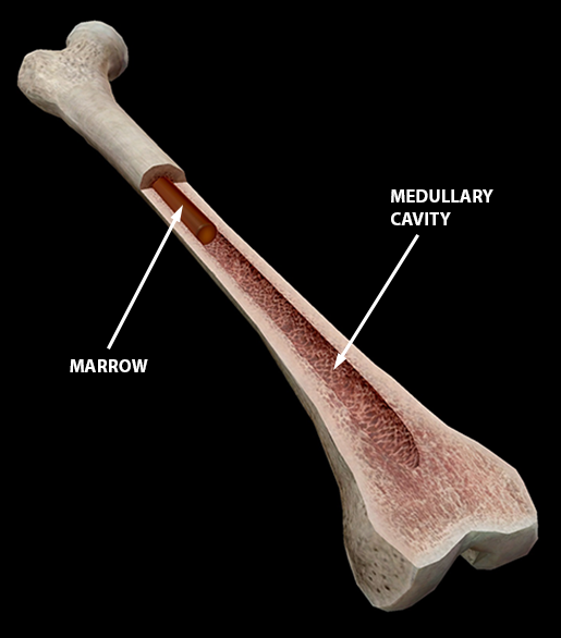
The structure of a long bone allows for the best visualization of all of the parts of a bone (figure 1). The hollow region in the diaphysis is called the medullary cavity, which is filled with yellow. If the cause is large or complex, it is best to break it down into sub causes. The end of a long bone. Female pelvic bone anatomy images. A long bone is a bone that has greater length than width. Learn long bone with free interactive flashcards. Inside the diaphysis is the medullary cavity, which is filled with yellow bone marrow in an adult. The calf bone or fibula is the smaller of the two bones that form the lower leg. Diagram of a radious bone 12 photos of the diagram of a radious bone diagram of radius bone, bone, diagram of radius bone. A hollow medullary cavity is found in the center of long bones and serves as a storage area for bone marrow. In this step, you will possibly have the diagram in front of you. The diaphysis is the tubular shaft that runs between the proximal and distal ends of the bone.
Komentar
Posting Komentar